October 30, 2017. CytoGnomix signs agreement with International Atomic Energy Agency

Cytognomix logo

NEWS RELEASE

For immediate release

Ontario company contributes to radiation biodosimetry project at the International Atomic Energy Agency

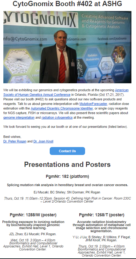
Cytognomix accelerates estimation of radiation exposure by participating institutions of International Atomic Energy Agency (IAEA) Member States

October 30, 2017                                London, Ontario, Canada                              Cytognomix Inc

Calibration of radiation exposure needs to be accurate for effective cancer treatment. Treatment of radiation overexposures depends on precise measurement of absorbed dose and the type of radiation received. Quantification of radiation exposure by biodosimetry testing needs to be timely for patients to benefit.

The IAEA is committed to encouraging and assisting research on, and development of practical applications of atomic energy for peaceful uses throughout the world. It has extended the opportunity to research institutes in Member States to participate in the Coordinated Research Project (CRP) E35010 entitled ‘Applications of Biological Dosimetry Methods in Radiation Oncology, Nuclear Medicine, and Diagnostic and Interventional Radiology.’

The IAEA is sponsoring CytoGnomix’s Research Project, entitled ‘Determination of Radiation Exposure by Fully Automated Dicentric Chromosome Analysis.’  This project will enable laboratories and research institutions of Member States to use Cytognomix’s technology to accelerate testing radiation exposure.

In this project, Cytognomix Inc. will use its systems to automatically analyze digital images of chromosomes exposed to radiation to estimate exposure. Results obtained by biodosimetry laboratories at Health Canada and Canadian Nuclear Laboratories suggest that the results are similar to traditional manual analyses, but are achieved considerably more quickly. This research will expand access to these systems by other laboratories participating in IAEA’s Coordinated Research Project.

Accuracy and speed of the automated system  will be compared with previous results from collaborating CRP laboratories that were obtained by manual or computer-assisted DCA scoring. It is  anticipated that cell image data obtained from test samples in prior or current international joint laboratory exercises or  independent assay validation activities will be reused in this study. Each collaborating laboratory will also receive a demonstration software version containing their calibration curve and test sample data. Dose estimates obtained by CytoGnomix will be  compared with results obtained by collaborators. If the previous results are comparable to those obtained with ADCI in  different laboratories, this will establish the feasibility of undertaking larger scale, batch analysis of populations of individuals that  have potentially received radiation exposure.  A unique aspect of the proposed study will assess whether it enables greater standardization of results obtained by  different laboratories, because all labs will use a common algorithm to process their data, while still allowing different labs to  customize their own calibration curves for determining unknown radiation exposures, which addresses differences in chromosome preparation methods and radiation calibration sources between labs.

Quotes

“The IAEA has recognized the critical need for faster approaches to accurately determine radiation exposure that address impending needs by its members. By sponsoring our project, CytoGnomix will have a unique opportunity to provide hands-on experiences to radiation biodosimetry laboratories and centres worldwide.”

Dr. Peter K. Rogan

President of Cytognomix Inc.


Quick facts

  • Established in 2009, CytoGnomix Inc. is a biotechnology company that designs and markets advanced genomic reagents and software-based solutions. Its products personalize the diagnosis, evaluation and management of cancer, prenatal disorders and other genetic diseases.
  • CytoGnomix’s ADCI software system selects high-quality cells from all types of digital images for analysis, identifies chromosome anomalies, builds biodosimetry calibration curves and estimates exposure in less than an hour.
  • In 2017, IAEA is sponsoring 35 Cooperative Research Activities on diverse topics concerning the peaceful use of atomic energy. CRP E35010, which is focused on the biological effects of radiation, is one of 5 projects focused on human health. The decision to award a research contract or agreement is made after careful consideration of the technical merits of the proposal, the compatibility of the project with the IAEA’s own functions and approved programmes, the availability of appropriate facilities and personnel in the institution and previous research work related to the project. Where it is recognized that the award of a particular research or technical contract or research agreement would materially assist one of the IAEA’s programmes, an invitation is sent to those institutions believed to have the necessary facilities and personnel, and the Government of the Member State concerned is kept informed.


Associated links

CytoGnomix: company website;  radiation biodosimetry website

Follow us on Facebook

IAEA:  website

Follow us on Facebook


Contacts

Corporate Communications, CytoGnomix

(1) 519-661-4255

info@cytognomix.com


October 4, 2017. Three upcoming presentations at the American Society of Human Genetics annual conference

PgmNr 182: Splicing mutation risk analysis in hereditary breast and ovarian cancer exomes. (Platform)

Thurs, Oct 19. 11:00am -12:30pm. Session 40. Defining High Risk in Cancer. Room 230C – Level 2/Orlando Convention Center 

E.J. Mucaki 1; B.C. Shirley 2; S.N. Dorman 1; P.K. Rogan 1,2  1) Biochemistry, University of Western Ontario, London, Ontario, Canada; 2) CytoGnomix Inc, London, Ontario, Canada


Genetic testing of patients with inherited cancer frequently reveals variants of unknown significance (VUS). We have presented an Information Theory (IT) framework to predict and prioritize coding and non-coding VUS in hereditary breast and ovarian cancer (BRCA) patients, including effects on mRNA splicing1,2. We investigated the exome wide distribution of predicted mRNA splicing mutations in a large BRCA cohort. Predicted splicing mutations in IT-based splicing analysis of all variant data from AmbryShare BRCA exome (n=11,416; with 1.2 million VUS) and the control genome Aggregation Databases (gnomAD; n=138,632) were identified using the Shannon splicing mutation software pipeline3. IT-flagged variant frequencies (decreasing Ri values [in bits] of either leaky or inactivated natural splice sites [∆Ri >4 bits and Ri ≤ 1.6] or strengthened cryptic splices sites with an Ri exceeding that of adjacent natural sites) were compared for each gene using odds ratios (OR). ORA is defined as the ratio of frequencies of the same flagged variants in a gene in AmbryShare relative to gnomAD. ORis based on the ratio of frequencies of all flagged variants in a gene in AmbryShare relative to all flagged variants in that gene in gnomAD. A greater number of IT-flagged variants were present in AmbryShare than in gnomAD among 2012 genes with severe splicing mutations. Increasing the ∆Ri threshold disproportionally decreases the number of flagged variants in gnomAD due to fewer severe splicing mutations. Variants that abolish natural splice sites flagged known inherited breast cancer genes with respectively increased ORand ORP inATM (493, 407), BARD1 (407, 407), BRCA1 (19, 14), BRCA2 (54, 54),CDH1 (549, 549), MLH1 (303, 303), MUTYH (95, 11), and PALB2 (233, 116). Other flagged breast cancer-related genes with high OR includeAAMP, C1QTNF6CDK3FOLR1PRLRRAD50RING1S100A2SRGN,TMSB10TYRO3, and VIM. Notable highly mutated genes from other cancers include GKN1 (gastric), C1orf61 (hepatocellular), CREM(prostate), PNKP (multiple), PPP1CA (gastric) and ZFAND2B (myeloid). Flagged genes not known to be linked to cancer include ATP1A4, MFF,PACSIN1PTS, and USH1C. Severe splicing mutations occur more frequently in inherited and somatic breast cancer genes as well as in other genes in BRCA populations.
1Mucaki et al. BMC Med. Genom. 9:19, 2016; 2Caminsky et al. Hum. Mut. 37:640, 2016; 3Shirley et al. Genom. Prot. Bioinf. 11:75, 2013.    Keywords: Cancer; Bioinformatics; Genomics; Population genetics; Statistical genetics


PgmNr 1268/T: Accurate radiation biodosimetry through automation of metaphase cell image selection and chromosome segmentation. (Poster)

Thurs, Oct 19.  2:00pm – 4:00pm. Bioinformatics and Computational Approaches. Exhibit Hall, Level 1, Orlando Convention Center 

Y. Li 1; J. Liu 2; B. Shirley 1; R. Wilkins 3; F. Flegal 4; J.H.M. Knoll 1,2; P.K. Rogan 1,2   1) CytoGnomix Inc, London, Ontario, Canada; 2) University of Western Ontario, London, Ontario Canada; 3) Health Canada, Ottawa, Ontario, Canada; 4) Canadian Nuclear Laboratories, Chalk River, Ontario, Canada


The dicentric chromosome (DC) assay is a standardized method that is recommended for determination of biologic radiation exposure1,2. Software to fully automate this assay has been developed in our laboratory3. This method relies on high quality microscope-derived images of metaphase cells to reduce the rate of false positive (FP) DCs. We present image processing methods to eliminate suboptimal metaphase cell images based on novel quality measures and to reclassify FPs by analyzing their morphological features. A set of chromosome segmentation thresholds selectively filtered out FPs, arising primarily from extended prometaphase chromosomes, sister chromatid separation and chromosome fragmentation. This reduced the number of FPs by 55% and was highly specific to the abnormal structures (≥97.7%). Image segmentation filters selectively remove images with consistently unparsable or incorrectly segmented chromosome morphologies, while image ranking sorts images according to their qualities and enables selection of optimal images in samples. Overall, these methods can eliminate at least half of the FPs detected by manual image review. By processing data to derive calibration curves and to assess samples of unknown exposures with the same image selection models, average dose estimation errors were reduced from 0.6 Gy to 0.3 Gy, without requiring manual review of DCs. During this presentation, we will use our software to demonstrate that metaphase image filtering and object selection constitute a reliable and scalable approach for biodosimetry, resulting in more accurate radiation dose estimates.

1. International Atomic Energy Agency. (2001) Cytogenetic Analysis for Radiation Dose Assessment, a Manual: Technical Reports Series. No. 405, International Atomic Energy Agency, Vienna.
2. International Atomic Energy Agency. (2011) Cytogenetic Dosimetry: Applications in Preparedness for and Response to Radiation Emergencies, International Atomic Energy Agency, Vienna.
3. Rogan, P. K., Li, Y., Wilkins, R. C., Flegal, F. N., and Knoll, J. H. M. (2016) Radiation Dose Estimation by Automated Cytogenetic Biodosimetry, Radiation Protection Dosimetry 172, 207-217.

Keywords: Bioinformatics; Centromere structure/function; Chromosomal abnormalities; Diagnostics; Public health

PgmNr 1288/W: Predicting exposure to ionizing radiation by biochemically-inspired genomic machine learning. (Poster)

Wed, Oct 18.  3:00pm – 4:00pm. Bioinformatics and Computational Approaches. Exhibit Hall, Level 1, Orlando Convention Center. 
J.Z.L. Zhao; E.J. Mucaki; P.K. Rogan.  Dept Biochemistry, University of Western Ontario, and CytoGnomix Inc., London, Ontario, Canada


Analyzing gene expression in peripheral blood mononuclear cells reveals profiles that predict radiation exposure in humans and mice by logistic regression (PLoS Med. 4:e106; PLoS ONE. 3:e1912). Using biochemically-inspired methods (Mol. Onc. 10:85-100), we derive gene signatures to predict the level of radiation exposure with improved accuracies. DNA repair genes responsive or differentially expressed upon radiation exposure and orthologs highly expressed in species resilient to radiation exposure (n=998) were analyzed by two-sampled t-tests comparing expression in individuals unexposed and exposed to radiation (150-200 cGy: humans or 50-1000 cGy: mice). Significance thresholds for including a gene in developing a signature were adjusted based on radiation dose, from p < 0.01 (50 cGy) to < 1E-14 (1000 cGy), equivalent to ~10% of genes. Support Vector Machine (SVM) signatures were derived by backward feature selection (BFS) or minimum-redundancy-maximum-relevance (mRMR) and validated using leave-one-out cross validation (LOOCV) and external datasets. GEO datasets GSE6874 and GSE10640 were used for training and testing. Signatures derived by BFS from the human patients of GSE6874 (n=78) included α) GADD45A, GTF3A, TNFRSF4, XPC and β) ATR, GADD45A, GTF3A, IL2RB, MYC, NEIL2, RBM15, SERPINB1, XPC, which both distinguished irradiated from unirradiated individuals with 98% sensitivity and 100% specificity in LOOCV. Validating these signatures on the human patients of GSE10640 (n=71) confirms that α and β are both 92% sensitive and, respectively, 94% and 96% specific. mRMR found the 10 “best” genes from the murine samples of GSE10640 (n=104) to create a signature at each radiation dose; several genes were common among signatures. Signature δ (50 cGy) included PHLDA3, BAX, NBN, CCT3, CDKN1A, CCNG1, POLK, ERCC5, GCDH, and RAMP1. Signature ε (200 cGy) included PHLDA3, LIMD1, CCT3, BAX, MS4A1, GLIPR2, BLNK, BCAR3, CDKN1A, andTFAM. Signature ζ (1000 cGy) included CCT3, SUCLG2, EI24, CNBP, PHLDA3, TPST1, HEXB, FEN1, CDKN1A, and BLNK. When validated on the murine samples of GSE6874 (n=14), each signature correctly predicted the exposure status of all mice. Our approach produces signatures with higher accuracies in cross- and external validation datasets than prior logistic regression models, with significantly improved sensitivities in detecting radiation exposure in humans. This will be useful in identifying nearly all radiation-exposed individuals in a mass casualty.

Keywords: Bioinformatics; Diagnostics; Transcriptome; Computational tools; Hematopoietic system