September 9, 2022. Update on US Patent Application “Smart microscope system for radiation biodosimetry”

Our patent application, US Patent Application Serial Number 17/137,317,  has had all claims allowed by the US Patent and Trademark office. This application covers the method underlying our ADCI Radiation biodosimetry software system. New claims cover partial body exposures, which are typical in radiation therapy. In addition, this invention covers applications of the technology which do not require interaction with the microscope system software, and can be used as a standalone system. The patent should be issued within the next several months.

Poster presentation at ConRad 2021 on radiation biodosimetry with ADCI

On May 10, 2021, CytoGnomix is presenting a poster at ConRad 2021 (www.radiation-medicine.de) titled:

Demonstration of the Automated Dicentric Chromosome Identifier and Dose Estimator [ADCI] System in a Cloud-based, Online Environment.

From the abstract:

Interpretation of cytogenetic metaphase images and quantification of exposures remain labour intensive in radiation biodosimetry, despite computer-assisted dicentric chromosome (DC) recognition and strategies to share workloads among different biodosimetry laboratories.  ADCI processes the captured images to identify DCs, selects images, and quantifies radiation exposure.  This paper describes ADCI_Online, a secure web-streaming platform  on Amazon Web Services that can be accessed worldwide from distributed local nodes.

ADCI_Online offers a subscription-based service useful for radiation research, biodosimetry proficiency testing, inter-laboratory comparisons, and training. In a research context, the system could provide highly uniform, reproducible assessment in large studies of many individuals, for example, exposed to therapeutic radiation. ADCI_Online compute environments originate from a single snapshot which can be cloned any number of times; thus, the system can be rapidly scaled when required. With robust network connectivity in a medical emergency of multiple potentially radiation exposed individuals, throughput and capacity for multiple samples requiring simultaneous processing and dose evaluation can be expanded to seamlessly mitigate any backlog in sample interpretation.

Platform presentation at ConRad 2021 on radiation gene signatures

On May 10, 2021, Dr. Rogan is giving a platform presentation at ConRad 2021 (www.radiation-medicine.de) titled
“Radiation biodosimetry exposure assessment from gene expression signatures can be confounded by other underlying disease pathologies.”

Misclassification of patients with underlying disorders by otherwise accurate radiation gene signatures compromises their utility for population-scale radiation exposure assessment. Underlying conditions modify the normal baseline values of biomarkers used for diagnostic analysis of radiation exposure. The collective frequency of these conditions would confound efforts to assess radiation exposures in a mass casualty event, affecting determination of eligibility for radiation-mitigating therapies.

#radiation #misdiagnosis #geneexpression #signature #medicine

September 4, 2020. New article on automated partial body radiation exposure determination

We have added the capability to determine whether samples exposed to ionizing radiation are wholly or partially irradiated. If partially, the approach determines the fraction of metaphase cells exposed and the whole body-equivalent dose completely automatically. CytoGnomix’s Automated Dicentric Chromosome Identifier and Dose Estimation software has been upgraded to generate these results as part of the the dose estimation report. The article has been accepted for publication by the International Journal of Radiation Biology  V. 96 (https://doi.org/10.1080/09553002.2020.1820611).  It is also currently available on BioRxiv:

Estimating partial body ionizing radiation exposure by automated cytogenetic biodosimetry
Ben C. ShirleyJoan H.M. KnollJayne MoquetElizabeth AinsburyPham Ngoc DuyFarrah NortonRuth C. WilkinsPeter K. Rogan

Full text:   ShirleyetalBioRxiv2020

January 9, 2019. Article on fully automated interpretation of the dicentric chromosome assay for radiation quantification now available

Our paper, “RADIATION DOSE ESTIMATION BY COMPLETELY AUTOMATED INTERPRETATION OF THE DICENTRIC CHROMOSOME ASSAY”  is now published in the journal Radiation Protection Dosimetry.

Unfortunately, the journal has not made the article open access. We have made it available on our ADCIWiki website, as permitted by the copyright agreement.

The link to the pdf full text is:

http://adcidewiki.cytognomix.com/files/pubs/LiRPD2019.pdf

January 28, 2013. Platform presentation on Automated Dicentric Chromosome Identifier Software

“Automating Dicentric Chromosome Detection from Cytogenetic Biodosimetry Data” at  the International EPRBioDose 2013 Conference in Leiden, Netherlands (March 24-28).

Authors: Peter Rogan(1,2), Akila Subasinghe(1), Asanka Wickramasinghe(1), Yanxin Li(1), Jagath Samarabandu(1), Joan Knoll(1,2), Ruth Wilkins(3), Farah Flegal(4); (1)University of Western Ontario, (2)Cytognomix Inc., (3)Health Canada, (4)Atomic Energy of Canada Ltd., Canada.

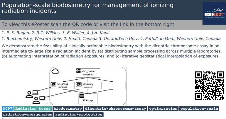
Abstract:  We are developing a prototype software system with sufficient capacity and speed to estimate radiation exposures by counting dicentric chromosomes in metaphase cells from many individuals in the event of a
mass casualty. Top-ranked metaphase images are segmented by defining chromosomes with an active contour gradient vector field (GVF), and by determining centromere locations along the centerline. The centerline is
extracted by Discrete Curve Evolution (DCE) skeleton branch pruning and curve interpolation. Centromere detection minimizes the global width and DAPI-staining intensity profiles along the centerline. A second
centromere is identified by reapplying this procedure after masking the first. Dicentrics can be identified by applying a support vector machine-based classification, which uses features that capture width and intensity
profile characteristics as well as local shape features of the object contour at candidate pixel locations. The correct location of the centromere is also refined in chromosomes with sister chromatid separation. The
overall algorithm has both high sensitivity (85%) and specificity (94%). Results are independent of the shape and structure of chromosomes in different cells, regardless of which laboratory protocol is followed or the
specimen source. The requisite throughput is being achieved by recoding MATLAB software modules for different segmentation functions in C++/OpenCV, and integrating them in the prototype. Processing of

numerous images is accelerated by both data and task software parallelization with the Message Passaging Interface and Intel Threading Building Blocks as well as an asynchronous non-blocking I/O strategy. Relative
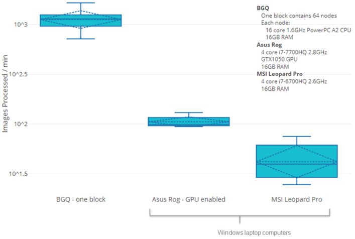
to a serial process, metaphase ranking, GVF, and DCE are respectively 100 and 300 fold faster on an 8-core I7-based desktop and on a 64-core shared memory cluster computer. Extrapolation from these benchmarks to
a 64-core system in which all of the software modules have been integrated indicates that it should be feasible to process metaphases for dicentric chromosomes from 1000 specimens in 20 hours.